Home — Essay Samples — Nursing & Health — Radiology — Nuclear Medicine

Nuclear Medicine
- Categories: Radiology
About this sample

Words: 379 |
Published: Mar 1, 2019
Words: 379 | Page: 1 | 2 min read
Works Cited
- International Atomic Energy Agency. (2019). Nuclear Medicine Resources Manual. Retrieved from https://www-pub.iaea.org/MTCD/Publications/PDF/Pub1777_web.pdf
- Ziessman, H. A., O'Malley, J. P., & Thrall, J. H. (2019). Nuclear Medicine: The Requisites. Elsevier.
- Fahey, F. H., & Zukotynski, K. (Eds.). (2016). Atlas of Nuclear Medicine. Springer.
- Henkin, R. E., Bova, D., Dillehay, G., & Jaszczak, R. (2019). Nuclear Medicine: A Core Review. Oxford University Press.
- Ell, P. J., Gambhir, S. S., & Hutton, B. F. (Eds.). (2019). Nuclear Medicine in Clinical Diagnosis and Treatment (4th ed.). Churchill Livingstone.
- O'Keefe, G. J., & Giammarile, F. (Eds.). (2016). Nuclear Medicine Therapy: Principles and Clinical Applications. Springer.
- Wong, K. K., Gould, M. K., & Wald, C. (Eds.). (2021). Nuclear Medicine: A Case-Based Approach. Springer.
- Saha, G. B. (2019). Fundamentals of Nuclear Pharmacy (7th ed.). Springer.
- Vallabhajosula, S. (Ed.). (2018). Molecular Imaging: Radiopharmaceuticals for PET and SPECT. Springer.
- Lin, E. C., & Buxbaum, S. G. (Eds.). (2020). Nuclear Medicine Physics: The Basics (9th ed.). Lippincott Williams & Wilkins.

Cite this Essay
To export a reference to this article please select a referencing style below:
Let us write you an essay from scratch
- 450+ experts on 30 subjects ready to help
- Custom essay delivered in as few as 3 hours
Get high-quality help

Prof Ernest (PhD)
Verified writer
- Expert in: Nursing & Health

+ 120 experts online
By clicking “Check Writers’ Offers”, you agree to our terms of service and privacy policy . We’ll occasionally send you promo and account related email
No need to pay just yet!
Related Essays
1 pages / 1435 words
2 pages / 812 words
2 pages / 1080 words
3 pages / 1705 words
Remember! This is just a sample.
You can get your custom paper by one of our expert writers.
121 writers online
Still can’t find what you need?
Browse our vast selection of original essay samples, each expertly formatted and styled
Related Essays on Radiology
Radiology, a field of medicine that utilizes various imaging techniques, has made remarkable advancements in recent years. This essay delves into the evolution of radiology technology, its diverse applications in diagnosing and [...]
Radiology and sonography are two essential diagnostic imaging techniques that play a crucial role in the field of medicine. Both methods utilize specialized equipment to produce images of the internal structures of the human [...]
Radiology, a field rooted in the visualization of the human body, has undergone a transformative journey with the integration of artificial intelligence (AI). This essay explores the burgeoning relationship between radiology and [...]
In my country, Bangladesh, there is a stereotypical conception that if a student doesn't accomplish his undergraduate degree from a renowned government academy, he is incompetent. But I always begged to differ and rather [...]
If the ability to cure a deadly and deliberating disease like diabetes is within a scientist’s grasps, should it be taken? What if this involves offering or copying genetic DNA to accomplish this task? Individuals within the [...]
There are a lot of innovations in medicine around the world that have many beneficial effects for humanity. States like Japan, Germany, Italy are known for such great medical innovations. But, United States remains the world [...]
Related Topics
By clicking “Send”, you agree to our Terms of service and Privacy statement . We will occasionally send you account related emails.
Where do you want us to send this sample?
By clicking “Continue”, you agree to our terms of service and privacy policy.
Be careful. This essay is not unique
This essay was donated by a student and is likely to have been used and submitted before
Download this Sample
Free samples may contain mistakes and not unique parts
Sorry, we could not paraphrase this essay. Our professional writers can rewrite it and get you a unique paper.
Please check your inbox.
We can write you a custom essay that will follow your exact instructions and meet the deadlines. Let's fix your grades together!
Get Your Personalized Essay in 3 Hours or Less!
We use cookies to personalyze your web-site experience. By continuing we’ll assume you board with our cookie policy .
- Instructions Followed To The Letter
- Deadlines Met At Every Stage
- Unique And Plagiarism Free
- COP Climate Change
- Coronavirus (COVID-19)
- Cancer Research
- Diseases & Conditions
- Mental Health
- Women’s Health
- Circular Economy
- Sustainable Development
- Agriculture
- Research & Innovation
- Digital Transformation
- Publications
- Academic Articles
- Health & Social Care
- Environment
- HR & Training
- Stakeholders
- Asia Analysis
- North America Analysis
- Our Audience
- Marketing Information Pack
- Prestige Contributors
- Testimonials

- North America
- Open Access News
- Research & Innovation News
The role of nuclear medicine in modern society
Ag spoke to dr arturo chiti, president elect of the european association for nuclear medicine (eanm) about the role nuclear medicine plays in modern society, and its challenges..
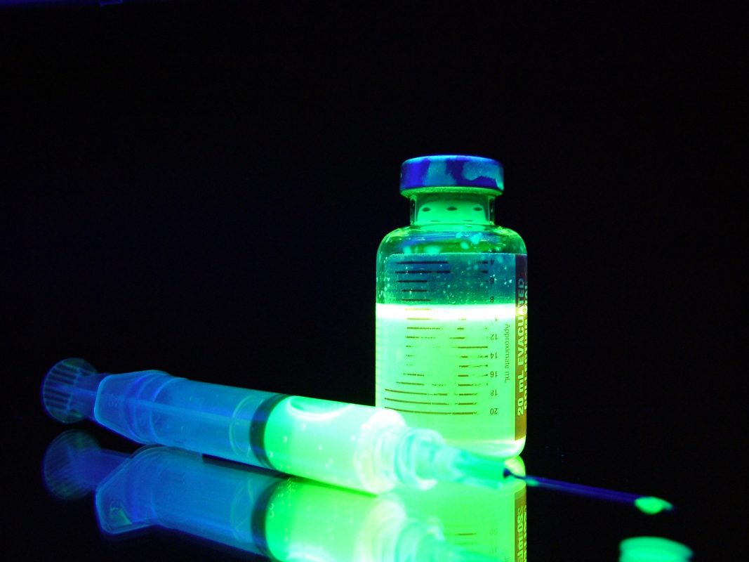
In modern society there are a number of healthcare challenges that researchers are fighting against to find new prevention methods and treatments for. Diseases such as cancer can be treated more successfully if caught and diagnosis is made much earlier.
Nuclear medicine plays a pivotal role in this and can help give medical staff the early diagnosis for the patient, and a more localised point for treatment. The European Association of Nuclear Medicine (EANM) works to educate people in the method of nuclear medicine, and further advance methods to help with that all important early diagnosis. However, when people hear the word nuclear, it can generate a fear in some, and without the correct information patients don’t understand how nuclear medicine can help prevent and treat some of those major health challenges.
In order to help give patients more information about molecular imaging and nuclear medicine techniques, The EANM have put together information for patients to remove the fear regarding this form of treatment, and set their minds at ease. Editor Laura Evans spoke to Doctor Arturo Chiti, President Elect of the EANM about how nuclear medicine fits into modern society, and the challenges that come with such a complicated field.
“One of the advantages is that nuclear medicine is able to see molecular alterations – what we call functional alterations in cancer,” explains Doctor Chiti.
“From research we have learnt that these alterations normally proceed the multi-functional alterations that we see with a CT scan. This is important because when you suspect cancer is present it helps in giving a very early diagnosis compared to morphological imaging.”
The method of nuclear medicine can be quite complicated, but it can help a number of healthcare problems from cancer to thyroid problems. Doctor Chiti explained the process, and the advantages this has with diseases such as cancer.
“Nuclear medicine or molecular imaging uses small probes that are called tracers, and these probes are able to localise particular tissues within the human body.
“These probes help to visualise diseases like cancer, or even evaluate how the blood flow goes into the heart. With this principle we are able to treat diseases like cancer, because these probes have radio-nuclides imbedded into the molecule, and radio-nuclides help you to localise exactly where the problem may be in order to treat it,” he says.
“Nuclear medicine helps us to do what we call personalised or precise medicine, because we can visualise the target and we can treat accordingly to this.”
Over the years nuclear medicine has evolved to keep up with modern medicine, and the constant health challenges faced throughout Europe. There are a number of ways in which this has happened, as explained by Doctor Chiti.
“We have 2 main tracks, the most important is the research of radiopharmaceuticals. These are the probes that are used in the process of nuclear medicine,” he says.
“In an effort to get more specific molecules to visualise or treat specific targets, we are carrying out research of radiopharmaceuticals for diagnosis, biological characterisation, and therapy. We can also design molecules which are exactly the same or similar to drugs which are used – non radioactive drugs.
“So the key aim is to be able to visualise the targets of drugs, which are used in oncology. This means you can select those patients that are going to benefit from a particular treatment.”
Technology also plays a vital role in molecular imaging, and one of the main challenges that Doctor Chiti pointed out was keeping up to date with sophisticated technology, as it evolves.
“We are using big hardware in order to track the radiopharmaceuticals which are injected into the patient, and this means that from that point of view we are aiming at having more and more sophisticated technology in order to visualise very small alterations in the human body,” says Doctor Chiti.
“Of course the imaging we use is always multi-modality imaging, that means that you have the molecular imaging, but you always have a CT scan or an MR scan as a companion in order to be able to have morphological and functional imaging in the patient at the same time. This increases the accuracy of the diagnoses we can do.”
Radiopharmaceuticals are the core of Doctor Chiti’s discipline, and it is the regulations relating to this area that he believes are one of the main challenges that faces the nuclear medicine field, as he explains.
“Regulatory issues are challenging because every radiopharmaceutical has to be approved at a European level, and then at a national level. Sometimes this is cumbersome and quite slow.
“Another issue is that radiopharmaceuticals are not developed at the same level throughout Europe. There are some countries where they are further developed than others. So, for example, there might be in country A, radiopharmaceutical for diagnosis or therapy available, but in neighbouring country B it is not. What we are aiming for is harmonised regulations throughout Europe – at least for radiopharmaceuticals.”
Looking ahead, Doctor Chiti would like to see nuclear medicine integrated with other clinical departments, in order to gain the best possible outcomes for patients.
“It will mean integrating nuclear medicine procedures with the other diagnostic imaging procedures – multi-modality imaging, and be more integrated in the oncological tracts, cardiological and neurological and also in those clinical tracts which are related to infection imaging,” he concludes.
“In my mind there will be more clinical speciality within this field of medicine, with more technology specialists working with the medical doctors.”
Doctor Arturo Chiti
President Elect
European Association of Nuclear Medicine (EANM)
Tel: +43 (0)1 212 80 30
www.eanm.org
RELATED ARTICLES MORE FROM AUTHOR

They weren’t witches; they were women: The witch-hunts and their repercussions

Webb telescope reveals cosmic question mark in early universe
Restoring learning ability in ageing brains

UK universities collaborate on revolutionary lightweight solar cells for space applications

James Webb Space Telescope reveals mysterious rogue worlds

What does your favourite film genre say about you?
Leave a reply cancel reply.
Save my name, email, and website in this browser for the next time I comment.
Related Academic Articles

Floreon technology, redefining polylactic acid

Fusion propulsion for exploring the solar system and beyond

Protecting the human epigenome with nutritional epigenetics intervention programs
Follow open access government, latest publication.

Open Access Government July 2024
- Terms & Conditions
- Privacy Policy
- GDPR Privacy Policy
- Marketing Info Pack
- Fee Schedule
What is Nuclear Medicine?
- Download PDF Copy

Nuclear medicine is the branch of medicine that involves the administration of radioactive substances in order to diagnose and treat disease. The scans performed in nuclear medicine are carried out by a radiographer. This speciality of nuclear medicine is sometimes referred to as endoradiology because the radiation emitted from inside the body is detected rather than being applied externally, as with an X-ray procedure, for example.
For nuclear medicine scans, radionuclides are combined with other chemical compounds to form the radiopharmaceuticals that are widely used in this field. When administered to the patient, these radiopharmaceuticals target specific organs or cellular receptors and bind to them selectively. External detectors are used to capture the radiation emitted from the radiopharmaceutical as it moves through the body and this is used to generate an image. Diagnosis is based on the way the body is known to handle substances in the health state and disease state.
The radionuclide used is usually bound to a specific complex (tracer) that is known to act in a particular way in the body. When disease is present, the tracer may be distributed or processed in a different way to when no disease is present. Increased physiological function that may occur as a result of disease or injury usually results in an increased concentration of the tracer, which can often be detected as a “hot spot.” Sometimes, the disease process leads to exclusion of the tracer and a “cold spot” is detected instead. A large variety of tracer complexes are used in nuclear medicine to visualize and treat the different organs, tissues and physiological systems in the body.
The main difference between nuclear medicine diagnostic tests and other imaging modalities is that nuclear imaging techniques show the physiological function of the tissue or organ being investigated, while traditional imaging systems such as computed tomography (CT scan) and magnetic resonance imaging (MRI scans) show only the anatomy or structure.
Nuclear medicine imaging techniques are also organ- or tissue-specific. While a CT or MRI scan can be used to visualize the whole of the chest cavity or abdominal cavity, for example, nuclear imaging techniques are used to view specific organs such as the lungs, heart or brain. Nuclear medicine studies can also be whole-body based, if the agent used targets specific cellular receptors or functions. Examples of these techniques include the whole-body PET scan or PET/CT scan, the meta iodobenzylguanidine (MIBG) scan, the octreotide scans, the indium white blood cell scan, and the gallium scan.
- http://unm.lf1.cuni.cz/vyuka/nuclear_medicine_jwfrank.pdf
- www.umich.edu/.../Sorenson_chpt-16.pdf
- https://www.uwlax.edu/
- https://www.snmmi.org/
- http://www.nupecc.org/pub/npmed2014.pdf
Further Reading
- All Nuclear Medicine Content
- History of Nuclear Medicine
- Nuclear Medicine Analysis
- Radiation Dose
- Jobs in Nuclear Medicine
Last Updated: Jun 12, 2023

Dr. Ananya Mandal
Dr. Ananya Mandal is a doctor by profession, lecturer by vocation and a medical writer by passion. She specialized in Clinical Pharmacology after her bachelor's (MBBS). For her, health communication is not just writing complicated reviews for professionals but making medical knowledge understandable and available to the general public as well.
Please use one of the following formats to cite this article in your essay, paper or report:
Mandal, Ananya. (2023, June 12). What is Nuclear Medicine?. News-Medical. Retrieved on September 08, 2024 from https://www.news-medical.net/health/What-is-Nuclear-Medicine.aspx.
Mandal, Ananya. "What is Nuclear Medicine?". News-Medical . 08 September 2024. <https://www.news-medical.net/health/What-is-Nuclear-Medicine.aspx>.
Mandal, Ananya. "What is Nuclear Medicine?". News-Medical. https://www.news-medical.net/health/What-is-Nuclear-Medicine.aspx. (accessed September 08, 2024).
Mandal, Ananya. 2023. What is Nuclear Medicine? . News-Medical, viewed 08 September 2024, https://www.news-medical.net/health/What-is-Nuclear-Medicine.aspx.
Suggested Reading

Cancel reply to comment
- Trending Stories
- Latest Interviews
- Top Health Articles

How can microdialysis benefit drug development
Ilona Vuist
In this interview, discover how Charles River uses the power of microdialysis for drug development as well as CNS therapeutics.

Global and Local Efforts to Take Action Against Hepatitis
Lindsey Hiebert and James Amugsi
In this interview, we explore global and local efforts to combat viral hepatitis with Lindsey Hiebert, Deputy Director of the Coalition for Global Hepatitis Elimination (CGHE), and James Amugsi, a Mandela Washington Fellow and Physician Assistant at Sandema Hospital in Ghana. Together, they provide valuable insights into the challenges, successes, and the importance of partnerships in the fight against hepatitis.

Addressing Important Cardiac Biology Questions with Shotgun Top-Down Proteomics
In this interview conducted at Pittcon 2024, we spoke to Professor John Yates about capturing cardiomyocyte cell-to-cell heterogeneity via shotgun top-down proteomics.

Latest News

Newsletters you may be interested in
Your AI Powered Scientific Assistant
Hi, I'm Azthena, you can trust me to find commercial scientific answers from News-Medical.net.
A few things you need to know before we start. Please read and accept to continue.
- Use of “Azthena” is subject to the terms and conditions of use as set out by OpenAI .
- Content provided on any AZoNetwork sites are subject to the site Terms & Conditions and Privacy Policy .
- Large Language Models can make mistakes. Consider checking important information.
Great. Ask your question.
Azthena may occasionally provide inaccurate responses. Read the full terms .
While we only use edited and approved content for Azthena answers, it may on occasions provide incorrect responses. Please confirm any data provided with the related suppliers or authors. We do not provide medical advice, if you search for medical information you must always consult a medical professional before acting on any information provided.
Your questions, but not your email details will be shared with OpenAI and retained for 30 days in accordance with their privacy principles.
Please do not ask questions that use sensitive or confidential information.
Read the full Terms & Conditions .
Provide Feedback

Nuclear Medicine and Molecular Imaging Essay
- To find inspiration for your paper and overcome writer’s block
- As a source of information (ensure proper referencing)
- As a template for you assignment
In current healthcare settings, nuclear medicine has become an important tool for aiding patients. Despite the dangers associated with the use of radioactive elements, through their application in diagnostics and treatment, it is possible to limit their harm and enhance their benefits. In particular, these types of tools are invaluable when working on treating various types of cancer. For example, lymphoma, as the type of cancer that affects the body’s blood system, usually requires the use of nuclear medicine. Before any treatment is administered, molecular imaging is used to determine the diagnosis and the severity of the disease. Compared to other types of imaging, it enables doctors and physicians to examine patients on the deepest of levels and detect the presence of cancer and other disturbances (“Fact sheet: Molecular imaging and lymphoma,” n.d.). From that point onward, nuclear medicine serves as a tool for healing or managing the patient’s condition. Both chemotherapy and radiation therapy are widely used. These methods utilize either medicine or energy beams that are capable of attacking the cancer cells or managing their spread. Naturally, even these types of treatment are not always effective, and the ability of doctors to use them hinges on the patient’s response. However, it is undeniable that the application of radioactive and nuclear elements gives individuals more opportunities to live fulfilling lives.
Throughout this course, I was able to learn about a number of different treatment and diagnostics methods, as well as understand the development of the medical sphere in the past decades. I think that this knowledge gave me a newfound appreciation for researchers and doctors that strove to improve the tools at their disposal and create approaches capable of fighting even chronic diseases. In terms of my own everyday life, this knowledge helps me understand that concepts like harm or benefit are not always black and white, and many things that are inherently dangerous can be used responsibly. This applies to both medicine and all other kinds of human activity.
Fact sheet: Molecular imaging and lymphoma . (n.d.). Society of Nuclear Medicine and Molecular Imaging (SNMMI). Web.
- Bacterial Meningitis Diagnostics
- Hydration Experiment: Boosting Energy and Well-being
- China’s Future Economic Potential Hinges on Its Productivity
- Viruses as a Cause of Cancer
- Rabia of Basra's and Hadewijch of Brabant's Poetry
- Ménière’s Syndrome and Its Treatment
- Diagnosing a Child With Upper Respiratory Infection
- The Misinformation Associated With Tuberculosis Diagnosis
- Melancholic State: Differential Diagnostic Assessment
- Cholera Disease: Diagnostics and Treatment
- Chicago (A-D)
- Chicago (N-B)
IvyPanda. (2024, February 29). Nuclear Medicine and Molecular Imaging. https://ivypanda.com/essays/nuclear-medicine-and-molecular-imaging/
"Nuclear Medicine and Molecular Imaging." IvyPanda , 29 Feb. 2024, ivypanda.com/essays/nuclear-medicine-and-molecular-imaging/.
IvyPanda . (2024) 'Nuclear Medicine and Molecular Imaging'. 29 February.
IvyPanda . 2024. "Nuclear Medicine and Molecular Imaging." February 29, 2024. https://ivypanda.com/essays/nuclear-medicine-and-molecular-imaging/.
1. IvyPanda . "Nuclear Medicine and Molecular Imaging." February 29, 2024. https://ivypanda.com/essays/nuclear-medicine-and-molecular-imaging/.
Bibliography
IvyPanda . "Nuclear Medicine and Molecular Imaging." February 29, 2024. https://ivypanda.com/essays/nuclear-medicine-and-molecular-imaging/.
Advertisement
Nuclear medicine: a global perspective
- Published: 25 March 2020
- Volume 8 , pages 51–53, ( 2020 )
Cite this article

- Diana Paez 1 ,
- Francesco Giammarile 1 &
- Pilar Orellana 1
8803 Accesses
16 Citations
3 Altmetric
Explore all metrics
Avoid common mistakes on your manuscript.
In this era of globalization, health systems, primarily in low- and middle-income countries (LMICs) [ 1 ] are required to accommodate the care and management of the increasing burden of non-communicable diseases (NCDs). Vast regions of the world also must contend with the double burden of disease, including facing outbreaks of novel viruses. Health systems need to address the epidemiological transition from communicable to NCDs with strengthened capacity. Nuclear Medicine has become an integral part of an efficient health system that acknowledges this shift with the management of various NCDs, in particular cardiovascular, oncological, and neurodegenerative diseases.
Without a doubt, innovation in research and development is a driving force in nuclear medicine. New devices, radiopharmaceuticals, clinical applications, and evidence-based medicine are produced at a fast pace and need to be propagated. New standards of best practices should be emphasized not only as part of training programmes, but also all efforts should be made to keep the medical community abreast with developments to provide optimal patient care and professional growth.
Global health realities show large inequities in access to nuclear medicine services. These are now showcased in an unprecedented, comprehensive manner in the NUclear mEdicine DAtaBase (NUMDAB) [ 2 ], as well as in the IAEA Medical imAGIng and Nuclear mEdicine global resources database (IMAGINE) [ 3 ].
According to the data available at International Atomic Energy Agency (IAEA) [ 2 , 3 ], 134 out of 195 countries have nuclear medicine facilities. There are over 25,000 SPECT cameras. The heterogenous global distribution of SPECT systems range between 0.036 cameras/million inhabitants in low-income countries (LIC) to 17.9 cameras/million in high-income countries (HIC). It should also be noted that the difference between upper middle-income countries (UMIC) and lower middle-income (LMIC) countries is sixfold (1.33 cameras/million vs 0.20 cameras/million, respectively) (Table 1 ). The regional distribution is also heterogenous. In the Americas, the USA has the highest rate of SPECT cameras/million inhabitants (45.3), whereas Canada has 16 cameras/million. There are 1456 cameras, with a rate of 3.73 cameras/million in Latin America and the Caribbean (LAC). Ranging from Argentina with 8.74 cameras/million, Brazil with 6.36, Colombia with 2.8 cameras/million inhabitants, Ecuador with 0.7 scanners/million to countries such as Aruba or Barbados with no availability of SPECT scanners.
Inequities in access to PET-CT are more striking. In high-income countries, there are 3.2 scanners/million inhabitants and in low-income settings, 0.007 scanners/million (Table 1 ).
Regionally, using the African continent as an example, only 9 out of 54 countries have PET scanners.
Stimulating market projections to inject investment and research and development is key to maintaining and growing nuclear medicine access. Currently, the discipline of diagnostic nuclear medicine (SPECT and PET) has the smallest share of the global medical imaging market (including CT, MRI, US, and X-ray) at 6.5%. It is expected to be the fastest growing segment due to the rising prevalence of NCDs, increased needs for early and accurate diagnoses, new technological developments both in hardware and software, the availability of new tracers, and its welcomed reception in emerging markets.
The LAC region serves to illustrate the global nuclear medicine growth. The practice of nuclear medicine in the region has experienced an important development in the last decades. However, there is great heterogeneity among countries regarding the availability of technology and human resources. According to data collected by the IAEA throughout the years, PET scanners in the LAC region had a Compound Annual Growth Rate of approximately 21% and grew from 22 systems in 6 countries (out of the 33 countries in the region) in 2005, to 144 systems in 11 countries in 2015, and 301 systems in 17 countries in 2019. Although the growth was higher than the global nuclear medicine imaging devices market, the number of PET scanners per million inhabitants is around 0.47. In the Middle East (ME), the number of PET scanners in 2016 was 194 [ 4 ] and grew to 220 scanners in 2019, the number of PET scanners per million inhabitants in the ME is 0.66 [ 3 ]. In both cases LAC and the ME, the number of PET scanners/million inhabitants is still far below the recommended 2.0 to 2.5 scanners per million in an optimal health setting [ 5 ]. As for the SPECT market, there existed 1200 systems in 2015 [ 6 ] in 20 out of the 33 LAC countries to 1456 systems in 2019, distributed in 23 countries [ 3 ].
In this era of unprecedented scientific and technological developments there is an increasing demand for “a personalized medicine approach” which is the development of better strategies for detecting and treating diseases based on an individual’s unique profile. The growth of personalized medicine is propelled by several factors that include the advances in molecular biology, genetics and proteomics, the better understanding of normal and pathological processes, the greater knowledge of the mechanism of individual diseases, the superior identification of disease subtypes and the better prediction of individual patient’s responses to treatment.
Expanded use of nuclear medicine techniques has the potential to accelerate, simplify, and reduce the costs of developing and delivering improved health care, reduce the healthcare expenditure and could facilitate the implementation of personalized medicine. Its applications can go far beyond diagnostics, allowing support in the selection of the appropriate therapy, evaluating the therapy response and follow-up; and driving the journey to personalized medicine.
Nuclear medicine has come to a fork in the road, with the choice being whether to remain a diagnostic lesion-detection technique, or whether to join the revolution in precision medicine, characterizing tumour biology and directing treatment through highly specific tracers that guide us to the world of theragnostic with translation molecular imaging being the central focus (Fig. 1 ).
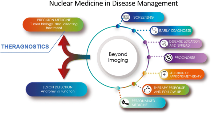
Nuclear medicine in disease management
To reach the full potential of nuclear medicine, including theragnostic, it is essential to train nuclear medicine professionals, not only newcomers but also experienced professionals trained in the field; to work with regulatory bodies to ensure and comply with safety requirements; to cooperate alongside policy makers, to include reimbursements for established and emerging applications, and to implement quality management systems in clinical practice to ensure the safety and quality of services provided.
Nuclear medicine and medical imaging should be included in the main global public policy guidelines for management of patients. Multisectoral coordination amongst key stakeholders (World Health Organization, IAEA, and other United Nations organizations; governmental bodies, professional organizations, non-governmental organizations, and private sector) is crucial to be able to deliver the best care possible. Unfortunately, this multisectoral coordination is currently unclear.
There are some ongoing efforts by the international community to deal with the burden of diseases. In 2015, the United Nations established the Sustainable Development Goals, a set of 17 specific goals aimed at transforming our world by 2030 and achieving a better and more sustainable future for all [ 7 ]. Goal 3 focuses on ensuring healthy lives and promoting well-being for all at all ages. It is important to highlight two targets: target 3.4 addresses the challenges of reducing NCD premature mortality by 30% by 2030 and target 3.9 aims at strengthening the capacity of all countries, particularly developing countries, for early warning, risk reduction and management of national and global health risks.
To support national efforts to address the burden on NCDs, the 66th World Health Assembly endorsed the WHO Global Action Plan (GAP) for prevention and control of NCDs 2013–2020 (resolution WHA66.10) [ 8 ]. The GAP offers a paradigm shift by providing a roadmap and policy options menu for Member States, the WHO, other UN organizations, intergovernmental organizations, NGOs and the private sector. The hypothesis is that, if implemented collectively between 2013 and 2020, we will achieve nine voluntary global targets, including a relative 25% reduction in premature NCDs mortality by 2025. Target 8 is of pivotal importance to the medical imaging community. Its objective is to achieve 80% availability of affordable basic technologies and essential medicines, including generics required to treat NCDs in both public and private facilities. The problem is that medical imaging and nuclear medicine are not defined under target 8. Nevertheless, there is an opportunity to include medical images as affordable or basic technologies, for which the professional community must participate in decision-making processes.
It is fundamental to advice the health authorities of the importance of including medical imaging and nuclear medicine as part of the policies, strategies, and action plans for health technologies and the national health plan.
World Bank Group. New Country Classifications by Income Level: 2019–2020. https://blogs.worldbank.org/opendata/new-country-classifications-income-level-2019-2020
NUMDAB, NUclear Medicine DAtaBase. https://humanhealth.iaea.org/HHW/NuclearMedicine/NUMDAB/index.html
IMAGINE, the new IAEA Medical imAGIng and Nuclear mEdicine global resources database. https://humanhealth.iaea.org/HHW/DBStatistics/IMAGINE.html
Paez D, Becic T, Bhonsle U, Jalilian AR, Nuñez-Miller R, Osso JA Jr (2016) Current status of nuclear medicine practice in the Middle East. Semin Nucl Med 46(4):265–272
Article Google Scholar
International Atomic Energy Agency, Planning a Clinical PET Centre, Human Health Series No. 11, IAEA, Vienna (2010)
Páez D, Orellana P, Gutiérrez C, Ramirez R, Mut F, Torres L (2015) Current status of nuclear medicine practice in Latin America and the Caribbean. J Nucl Med 56(10):1629–1634
United National Sustainable Development Goals. https://sustainabledevelopment.un.org
World Health Organization. Global Action Plan for Healthy Lives and Well-being for All. https://www.who.int/sdg/global-action-plan
Download references
Author information
Authors and affiliations.
Nuclear Medicine and Diagnostic Imaging Section, Division of Human Health, International Atomic Energy Agency, Vienna International Centre, PO Box 100, 1400, Vienna, Austria
Diana Paez, Francesco Giammarile & Pilar Orellana
You can also search for this author in PubMed Google Scholar

Contributions
All authors contributed equally to the paper.
Corresponding author
Correspondence to Francesco Giammarile .
Ethics declarations
Conflict of interest.
The authors declare that they have no conflict of interest.
Research involving human and animal rights
This article does not contain any studies with human participants or animals performed by any of the authors.
Informed consent
For this type of study, informed consent is not required.
Additional information
Publisher's note.
Springer Nature remains neutral with regard to jurisdictional claims in published maps and institutional affiliations.
Rights and permissions
Reprints and permissions
About this article
Paez, D., Giammarile, F. & Orellana, P. Nuclear medicine: a global perspective. Clin Transl Imaging 8 , 51–53 (2020). https://doi.org/10.1007/s40336-020-00359-z
Download citation
Published : 25 March 2020
Issue Date : April 2020
DOI : https://doi.org/10.1007/s40336-020-00359-z
Share this article
Anyone you share the following link with will be able to read this content:
Sorry, a shareable link is not currently available for this article.
Provided by the Springer Nature SharedIt content-sharing initiative
- Find a journal
- Publish with us
- Track your research
- Advanced search

Advanced Search
Nuclear Medicine: The Essentials
- Find this author on Google Scholar
- Find this author on PubMed
- Search for this author on this site
- For correspondence: [email protected]
- Info & Metrics
H. Jadvar and P.M. Colletti
Wolters Kluwer, 2021, 310 pages, $110.99
In contrast to the discipline of conventional radiology, to which medical school students, trainees, and many practitioners of medicine are heavily exposed, the field of nuclear medicine is somewhat specialized and requires special training for optimal understanding of its role in various domains in medicine. Therefore, there is a dire need for a simplified exposure to the specialty that provides some practical knowledge about the field and its unique role in the day-to-day practice of medicine. “The Essentials” series is a collection of radiology textbooks that follow such a standardized format. The series is designed to provide a practical tool for those who wish to gain a broad base of knowledge on various specialties in medical imaging. The content is confined to the essentials of the specialty and can be understood by the novice. However, enough details are included to be useful for those who teach the specialty and to provide a reference for health-care providers practicing the specialty of imaging. “The Essentials” books are compact in size and allow for residents and other interested groups to grasp practical knowledge about the various procedures that are offered by this specialty. Furthermore, the self-assessment sections provide multiple-choice questions at the end of each chapter. As such, this additional training is of particular benefit for those who are preparing for an image-rich computer-based examination for professional and maintenance certifications.
Currently, the field of nuclear medicine is the fastest-growing discipline in medical imaging. The recent introduction of novel radiopharmaceuticals for imaging and targeted therapy is revolutionary; therefore, educating trainees and the community at large about their applications in many disciplines is essential at this time. These include innovations in high-technology instruments related to digital and time-of-flight cameras, total-body PET instruments, PET/CT, PET/MRI, and SPECT/CT. This textbook provides a concise yet comprehensive overview of the field of molecular imaging that fits the criteria intended for “The Essentials” series. Each chapter describes the basics of physics, instrumentation, quality control, radiochemistry, radiation safety, and other essential information about each procedure.
The table of contents includes radiochemistry, instrumentation, physics, and radiation safety as introductions to technical bases for performing various procedures. The clinical section deals with assessment of diseases and disorders of various organs and anatomic structures (thyroid, parathyroid, and neuroendocrine glands; central nervous system; skeleton; lungs; gastrointestinal tract; kidneys; and lymph nodes). Also, chapters are devoted to radiotheranostics, the essentials of pediatric nuclear medicine, quality assurance, and procedures on pregnant and lactating patients. Overall, the book includes 19 chapters.
The chapters are organized in a logical manner and describe in some detail the imaging techniques that practitioners of the discipline follow. Therefore, readers who may not be familiar with the role of nuclear medicine procedures will be able to comprehend the scope of this discipline in clinical settings. No critically important topics are missing from this comprehensive book.
The chapters are written by highly qualified and expert contributing authors with longstanding experience in their respective disciplines. The main authors, Drs. Jadvar and Colletti, have substantially contributed by writing several chapters of this book.
Overall, this book provides a well-balanced view of current applications of conventional nuclear medicine and PET. Therefore, the book is a strong medium for introducing physicians and scientists to ongoing activities in the field and their relevance to the day-to-day practice of medicine. There are no serious weaknesses to the overall content of the book. Additionally, the figures and tables are of high quality. Most of the figures in the book are selected from the authors’ own clinical files and are of high quality.
In conclusion, Nuclear Medicine: The Essentials provides a comprehensive and excellent review of the current practice of the field. Therefore, this book will be of great interest to trainees, technologists, and scientists, as well as to practitioners of this rapidly evolving specialty. As such, the book is highly recommended for those who wish to refresh their understanding of the field and its various applications in medicine.
Published online Sep. 8, 2022.
- © 2023 by the Society of Nuclear Medicine and Molecular Imaging.
In this issue
- Table of Contents
- Table of Contents (PDF)
- About the Cover
- Index by author
- Complete Issue (PDF)
Thank you for your interest in spreading the word on Journal of Nuclear Medicine.
NOTE: We only request your email address so that the person you are recommending the page to knows that you wanted them to see it, and that it is not junk mail. We do not capture any email address.
Citation Manager Formats
- EndNote (tagged)
- EndNote 8 (xml)
- RefWorks Tagged
- Ref Manager

- Tweet Widget
- Facebook Like
- Google Plus One
Jump to section
Related articles.
- No related articles found.
- Google Scholar
Cited By...
- No citing articles found.
More in this TOC Section
- Magnetic Resonance Imaging in Movement Disorders: A Guide for Clinicians and Scientists
- Atlas of PET/MR Imaging in Oncology
Similar Articles
An official website of the United States government
The .gov means it’s official. Federal government websites often end in .gov or .mil. Before sharing sensitive information, make sure you’re on a federal government site.
The site is secure. The https:// ensures that you are connecting to the official website and that any information you provide is encrypted and transmitted securely.
- Publications
- Account settings
Preview improvements coming to the PMC website in October 2024. Learn More or Try it out now .
- Advanced Search
- Journal List
- Indian J Nucl Med
- v.33(3); Jul-Sep 2018
Dacryoscintigraphy: A Pictorial Essay
Shefali madhur gokhale.
Department of Nuclear Medicine, Inlaks and Budhrani Hospital, Pune, Maharashtra, India
Dacryoscintigraphy is a noninvasive, simple, easy to perform imaging modality used in the evaluation of epiphora. However, it is an infrequently done study in nuclear medicine departments. A standardized protocol and a systematic interpretation of the scans help in answering the queries of the clinicians in cases of epiphora. We have attempted to build a pictorial essay of the various findings detected on dacryoscintigraphy.
Introduction
Imaging the nasolacrimal system with use of radiopharmaceuticals, in other words, dacryoscintigraphy is an underutilized tool in the present-day nuclear medicine department.
Of the few nuclear medicine departments that perform the procedure, there are differences in the protocol followed, the use of collimators, and the use of radiopharmaceuticals. The indications for the procedure are evaluation of epiphora,[ 1 ] detection of subclinical lacrimal duct obstruction, appropriate patient selection for surgery,[ 2 ] and evaluating the success of dacryocystorhinostomy.[ 2 ] It is contraindicated in acute infective and allergic conditions of the eye. The small size of the structures of the nasolacrimal system is a major limitation. As compared to dacryocystography, the radiation exposure involved is about 100 times lower.[ 3 , 4 ] It has a distinct advantage over the syringing/saccharin test[ 5 ] in being noninvasive.[ 3 ] As there is no instrumentation of canaliculi or administration of contrast/saline under high pressure, false-negative and false-positive[ 3 , 5 ] results are avoided.
The standard protocol that we followed involved the use of Tc-99 m sulfur colloid in a dose of 50–100 microCi/10 μl[ 3 , 5 ] with the use of low-energy high-resolution collimator and images acquired immediately, postinstillation of normal saline drops and postblowing of the nose.[ 6 ]
A systematic interpretation of the various sequences helps arrive at the etiology of epiphora.
Dacryoscintigraphy interpretation in patients presenting with epiphora
To demonstrate normal flow through the nasolacrimal system.
A case of dacryocystorhinostomy on the left side in a 26-year-old male patient.
Drainage of tracer into the nasal cavity within the first 15-min postinstillation of radiopharmaceutical is considered normal.
The left eye reveals flow of tracer to the medial canthus region with accumulation of tracer there. There is no drainage of tracer into the nasal cavity.
The right eye reveals flow of tracer into the lacrimal sac, nasolacrimal duct (NLD), and drainage into the inferior meatus of the nose, all within the first 15-min postinstillation of Tc-99 m sulfur colloid, which is considered as normal [ Figure 1 ].[ 5 , 7 ]

Dacryoscintigraphy reveals drainage of tracer into the lacrimal sac (bold black arrow), nasolacrimal duct (hyphenated arrow), and inferior meatus of nose (black arrow) from the right eye. There is no drainage of tracer into the nasal cavity from the left eye
Interpretation
- Failed dacryocystorhinostomy on the left side
- Normal tear flow in the right eye.
To evaluate cause of epiphora in spite of a bilaterally normal syringing test
A 61-year-old female patient had a history of trauma to eyes approximately 2 months back. She had complaints of watering from the right eye for 2 months. Syringing revealed the passage of saline downward into the nasal cavity bilaterally.
The right eye reveals flow of tracer into the proximal NLD in immediate images. However, there is drainage into the inferior meatus of nose noted only in the images acquired postblowing of the nose.
The left eye reveals partial flow of tracer into the inferior meatus of the nose in the immediate images. There is persistent partial tracer stasis in the region of medical canthus noted in the delayed images [Figure [Figure2a 2a - -c c ].

(a) Immediate dynamic images of dacryoscintigraphy reveal drainage of tracer into the proximal nasolacrimal duct on the right side (bold black arrow). Partial drainage of tracer from the left eye into inferior meatus of the nose is noted (black arrow), (b and c) Dacryoscintigraphy images acquired postblowing of nose reveal flow of tracer from the right eye into inferior meatus of nose (bold black arrow). Persistent partial tracer stasis noted in the region of medial canthus of the left eye (black arrow)
Intraductal delay right eye with some inflammation at the lower end of NLD, which is relieved by nose blowing.
Partial functional impedance left eye.
The word “functional impedance” is used as an anatomic obstruction which is ruled out by the syringing test.[ 8 ] Hence, the finding of syringing test forms an important history in the interpretation of dacryoscintigraphy.
Epiphora since childhood
A 4.5-year-old female patient, a case of right-sided dacryocystitis. She has had complaints of right-sided epiphora since childhood.
Findings and Interpretation
The right eye reveals drainage of tracer into the NLD only in the image acquired postblowing of nose. This could be due to two reasons, either local inflammation at the NLD or resistance offered by the valves of the nasolacrimal system. Since this patient has had complaints since childhood, the symptoms are likely to be secondary to resistance offered by valves.
The left eye reveals drainage of tracer into the left NLD in immediate images. However, there is drainage into the inferior meatus of nose noted in the image acquired postblowing of nose. As the patient is asymptomatic on the left side, scan features are likely to represent local inflammation at the lower end of the left NLD [Figure [Figure3a 3a - -c c ]

(a) Immediate dynamic images of dacryoscintigraphy reveal flow of tracer into left nasolacrimal duct (black arrow), (b) there is no significant change in drainage observed after administration of normal saline drop, (c) images acquired after blowing of nose reveal flow of tracer into the nasolacrimal duct on the right (bold black arrow) and into the inferior nasal meatus on left (hyphenated arrow)
Bilateral epiphora, abnormal syringing test
A 64-year-old male patient with complaints of bilateral epiphora for 6–8 months.
There was no passage of saline detected on the syringing test bilaterally.
As there has been no passage of saline on the syringing test, there is likely to be an anatomic obstruction; however, the level of obstruction is to be detected.[ 5 ]
In the right eye, there is flow of tracer to the medial canthus region. However, the delayed images indicate that there is no flow of tracer into the lacrimal sac.
In the left eye, there is flow of tracer to the medial canthus region and lacrimal sac. However, there is no drainage into the NLD [Figure [Figure4a 4a - -c c ].

(a) Immediate dynamic images of dacryoscintigraphy reveal flow of tracer to the region of medial canthus bilaterally (arrows), (b) after administration of normal saline drops, no significant change in drainage of tracer is noted bilaterally, (c) delayed images reveal flow of tracer into the left lacrimal sac (arrowhead)
- PRESAC delay right eye
- PREDUCTAL delay left eye.
Epiphora in old age
A 59-year-old female diabetic and hypertensive patient presented with bilateral epiphora for 3–4 years.
There is pooling of tracer noted in the orbits bilaterally [Figure [Figure5a 5a and andb b ].

(a and b) Dacryoscintigraphy images reveal pooling of tracer in the orbits bilaterally (arrows)
This could be due to either failure of tear flow mechanism in the eyes or laxity of eyelids.
In this case, in view of the age of the patient and comorbidities, scan features are likely to be secondary to eyelid laxity.
To evaluate dacryocystorhinostomy on one side and epiphora on the other side
A 61-year-old male patient had presented with left lacrimal fossa abscess with dacryocystitis. At that time, syringing test had allowed passage of saline in the right eye. He underwent dacryocystorhinostomy on left side.
Findings and interpretation
The left eye reveals flow of tracer to the medial canthus region. However, there is no drainage of tracer into the left NLD. Hence, we conclude that the dacryocystorhinostomy on the left side has failed.
On the right side, we have an important clinical history of the syringing test allowing passage of saline. Hence, there is no anatomical obstruction noted on the right side.
Flow of tracer is noted to the right lacrimal sac. However, there is no drainage of tracer noted into the right NLD. This is a case of preductal delay. In view of the finding of syringing test, it is secondary to functional impedance on the right side [Figure [Figure6a 6a - -c c ].

(a) Dacryoscintigraphy images reveal drainage of tracer into the lacrimal sac bilaterally; however, there is no drainage of tracer into the nasolacrimal duct bilaterally. There is a minor difference in the radioactivity administered in both eyes, causing a minor difference in intensity of tracer to start with. As scan progresses, the difference in intensity of radiotracer in the two eyes increases as more tracer is flowing out of left eye and soaked out with tissue paper, (b) dacryoscintigraphy images reveal drainage of tracer into the lacrimal sac bilaterally; however there is no drainage of tracer into the nasolacrimal duct bilaterally, (c) dacryoscintigraphy images reveal no drainage of tracer into the nasolacrimal duct bilaterally
The word “impedance” is preferred by the ophthalmologists instead of “obstruction,” when the syringing test has allowed passage of saline.[ 8 ]
It is important to note that there is no transit of tracer into the NLDs bilaterally in patient V and patient VI. However, patient VI differs in having the flow of tracer to the medial canthus region on right side, hence changing the interpretation of the scans.
Dacryoscintigraphy is a simple noninvasive and physiological assessment of the nasolacrimal system. A standardized protocol and systematic interpretation would help us identify the cause of epiphora and ascertain the success of surgical procedures performed if any.
Declaration of patient consent
The authors certify that they have obtained all appropriate patient consent forms. In the form the patient(s) has/have given his/her/their consent for his/her/their images and other clinical information to be reported in the journal. The patients understand that their names and initials will not be published and due efforts will be made to conceal their identity, but anonymity cannot be guaranteed.
Financial support and sponsorship
Conflicts of interest.
There are no conflicts of interest.
An official website of the United States government
The .gov means it’s official. Federal government websites often end in .gov or .mil. Before sharing sensitive information, make sure you’re on a federal government site.
The site is secure. The https:// ensures that you are connecting to the official website and that any information you provide is encrypted and transmitted securely.
- Publications
- Account settings
- My Bibliography
- Collections
- Citation manager
Save citation to file
Email citation, add to collections.
- Create a new collection
- Add to an existing collection
Add to My Bibliography
Your saved search, create a file for external citation management software, your rss feed.
- Search in PubMed
- Search in NLM Catalog
- Add to Search
Dacryoscintigraphy: A Pictorial Essay
Affiliation.
- 1 Department of Nuclear Medicine, Inlaks and Budhrani Hospital, Pune, Maharashtra, India.
- PMID: 29962717
- PMCID: PMC6011558
- DOI: 10.4103/ijnm.IJNM_18_18
Dacryoscintigraphy is a noninvasive, simple, easy to perform imaging modality used in the evaluation of epiphora. However, it is an infrequently done study in nuclear medicine departments. A standardized protocol and a systematic interpretation of the scans help in answering the queries of the clinicians in cases of epiphora. We have attempted to build a pictorial essay of the various findings detected on dacryoscintigraphy.
Keywords: Dacryoscintigraphy; epiphora; nasolacrimal duct.
PubMed Disclaimer
Conflict of interest statement
There are no conflicts of interest.
Dacryoscintigraphy reveals drainage of tracer…
Dacryoscintigraphy reveals drainage of tracer into the lacrimal sac (bold black arrow), nasolacrimal…
(a) Immediate dynamic images of…
(a) Immediate dynamic images of dacryoscintigraphy reveal drainage of tracer into the proximal…
(a) Immediate dynamic images of dacryoscintigraphy reveal flow of tracer into left nasolacrimal…
(a) Immediate dynamic images of dacryoscintigraphy reveal flow of tracer to the region…
(a and b) Dacryoscintigraphy images…
(a and b) Dacryoscintigraphy images reveal pooling of tracer in the orbits bilaterally…
(a) Dacryoscintigraphy images reveal drainage…
(a) Dacryoscintigraphy images reveal drainage of tracer into the lacrimal sac bilaterally; however,…
Similar articles
- Dacryoscintigraphy: an effective tool in the evaluation of postoperative epiphora. Palaniswamy SS, Subramanyam P. Palaniswamy SS, et al. Nucl Med Commun. 2012 Mar;33(3):262-7. doi: 10.1097/MNM.0b013e32834f6cf7. Nucl Med Commun. 2012. PMID: 22186907
- The clinical value of dacryoscintigraphy in the selection of surgical approach for patients with functional lacrimal duct obstruction. Chung YA, Yoo IeR, Oum JS, Kim SH, Sohn HS, Chung SK. Chung YA, et al. Ann Nucl Med. 2005 Sep;19(6):479-83. doi: 10.1007/BF02985575. Ann Nucl Med. 2005. PMID: 16248384 Clinical Trial.
- Simultaneous dacryocystography and dacryoscintigraphy using SPECT/CT in the diagnosis of nasolacrimal duct obstruction. Kemeny-Beke A, Szabados L, Barna S, Varga J, Galuska L, Kettesy B, Gesztelyi R, Juhasz B, Toth L, Berta A, Garai I. Kemeny-Beke A, et al. Clin Nucl Med. 2012 Jun;37(6):609-10. doi: 10.1097/RLU.0b013e31824d2751. Clin Nucl Med. 2012. PMID: 22614201
- Lacrimal scintigraphy: "interpretation more art than science". Sagili S, Selva D, Malhotra R. Sagili S, et al. Orbit. 2012 Apr;31(2):77-85. doi: 10.3109/01676830.2011.648797. Orbit. 2012. PMID: 22489850 Review.
- Nuclear dacryoscintigraphy: its role in oral and maxillofacial surgery. Ziccardi VB, Charron M, Ochs MW, Braun TW. Ziccardi VB, et al. Oral Surg Oral Med Oral Pathol Oral Radiol Endod. 1995 Dec;80(6):645-9. doi: 10.1016/s1079-2104(05)80244-6. Oral Surg Oral Med Oral Pathol Oral Radiol Endod. 1995. PMID: 8680968 Review.
- Kemeny-Beke A, Szabados L, Barna S, Varga J, Galuska L, Kettesy B, et al. Simultaneous dacryocystography and dacryoscintigraphy using SPECT/CT in the diagnosis of nasolacrimal duct obstruction. Clin Nucl Med. 2012;37:609–10. - PubMed
- Chung YA, Yoo IeR, Oum JS, Kim SH, Sohn HS, Chung SK, et al. The clinical value of dacryoscintigraphy in the selection of surgical approach for patients with functional lacrimal duct obstruction. Ann Nucl Med. 2005;19:479–83. - PubMed
- Reddy SC, Zakaria A, Bhavaraju VM. Evaluation of lacrimal drainage system by radionuclide dacryoscintigraphy in patients with epiphora. Iran J Nucl Med. 2016;24:99–106.
- Kim HC, Cho AR, Lew H. Dacryoscintigraphic findings in the children with tearing. Korean J Ophthalmol. 2015;29:1–6. - PMC - PubMed
- MacDonald A, Burrell S. Infrequently performed studies in nuclear medicine: Part 1. J Nucl Med Technol. 2008;36:132–43. - PubMed
Related information
Linkout - more resources, full text sources.
- Europe PubMed Central
- Medknow Publications and Media Pvt Ltd
- Ovid Technologies, Inc.
- PubMed Central
- Citation Manager
NCBI Literature Resources
MeSH PMC Bookshelf Disclaimer
The PubMed wordmark and PubMed logo are registered trademarks of the U.S. Department of Health and Human Services (HHS). Unauthorized use of these marks is strictly prohibited.

IMAGES
VIDEO
COMMENTS
Nuclear medicine is a type of medical specialty that is used to diagnose and treat diseases using radioactive substances in a safe and painless way. It helps in determination of the medical conditions which is unavailable or require surgery or require very expensive diagnostic tests. It gives information of the molecular activity in the body ...
The Future of Nuclear Medicine, Molecular Imaging, and ...
Nuclear Medicine Essay. Nuclear Medicine is the use of radioisotopes for diagnosis, treatment, and research. Radioactive chemical tracers emit gamma rays which provides diagnostic information about a person's anatomy and the functioning of specific organs. Radioisotopes are also utilizes in treatments of diseases such as cancer.
Doctor Chiti explained the process, and the advantages this has with diseases such as cancer. "Nuclear medicine or molecular imaging uses small probes that are called tracers, and these probes are able to localise particular tissues within the human body. "These probes help to visualise diseases like cancer, or even evaluate how the blood ...
Nuclear Medicine. This chapter provides an overview of the field of nuclear medicine for readers who are not familiar with the discipline. It includes a description of the history and major discoveries in this field, the challenges of conducting nuclear medicine research, and the foreseeable new technologies and opportunities for personalizing ...
Citations. Nuclear medicine is the branch of medicine that involves the administration of radioactive substances in order to diagnose and treat disease. The scans performed in nuclear medicine are ...
Get a custom essay on Advanced Practice in Nuclear Medicine. Over the next 5-10 years, nuclear medicine has promising and various perspectives. As it was recently figured with cell-based therapies for prostate cancer (PSMA hitting CAR-T cells), more disorders could possibly be treated with radio therapeutical methods (Weber et al., 2020).
Nuclear medicine, a medical discipline focused on the use of radionuclides for the diagnosis and treatment of disease, traces its origins to the pioneering work of Henri Becquerel, Marie Skłodowska Curie, Pierre Curie, and George de Hevesy in the late 19th and early 20th centuries. 1-3 The idea of deploying synthetic radionuclides in medicine was first realised when John Lawrence explored ...
From that point onward, nuclear medicine serves as a tool for healing or managing the patient's condition. Both chemotherapy and radiation therapy are widely used. These methods utilize either medicine or energy beams that are capable of attacking the cancer cells or managing their spread.
The early years of nuclear medicine: A Retelling - PMC
Nuclear Medicine has become an integral part of an efficient health system that acknowledges this shift with the management of various NCDs, in particular cardiovascular, oncological, and neurodegenerative diseases. Without a doubt, innovation in research and development is a driving force in nuclear medicine. New devices, radiopharmaceuticals ...
It would take some 30 years before 3D Imaging became an integral part of the development of Nuclear Medicine. And one has to give proper due to a medical scientist, David Kuhl (Figure 3), the originator of single-photon emission tomography. Born in St. Louis, Missouri in 1929, David E. Kuhl graduated in medicine at UPENN in 1955.
Nuclear Medicine: The Essentials. Wolters Kluwer, 2021, 310 pages, $110.99. In contrast to the discipline of conventional radiology, to which medical school students, trainees, and many practitioners of medicine are heavily exposed, the field of nuclear medicine is somewhat specialized and requires special training for optimal understanding of ...
Nuclear medicine is a pharmaceutical that uses a small amount of radioactive material (radioisotopes) to diagnose, manage, treat and prevent diseases (i.e., cancer, cardiovascular diseases, neurological disorders, gastrointestinal, and other irregularities). This procedure can identify abnormal lesions within the body without the need for ...
601 Words. 3 Pages. Open Document. Nuclear Medicine. Argosy University. Nuclear medicine is a specialized branch of modern medicine that exploits the process of radioactivity for imaging, diagnosis, and treatment. Many imaging techniques inject small amounts of radioactive material into the body, which are then tracked by a sensing device ...
Nuclear Medicine The recent nuclear crisis suffered by Japan seems to justify the fear-mongering much of the mainstream media engages in regarding the use of nuclear energy. Yet, many experts know that "nuclear imaging is used on tens of thousands of patients every day to take pictures of their hearts, lungs, kidneys, bones, brains and other organs" (Walters, 2009, p.
The diagnosis and treatment of patients with cancer requires access to imaging to ensure accurate management decisions and optimal outcomes. Our global assessment of imaging and nuclear medicine resources identified substantial shortages in equipment and workforce, particularly in low-income and middle-income countries (LMICs). A microsimulation model of 11 cancers showed that the scale-up of ...
OBJECTIVE. The purpose of this pictorial essay is to illustrate several clinical situations in which SPECT/CT can be effectively applied in nuclear radiology practice. CONCLUSION. SPECT/CT has recently emerged as a valuable adjunct to standard techniques in clinical nuclear radiology. This technique provides significantly improved scintigraphic localization and characterization of disease ...
Dacryoscintigraphy is a noninvasive, simple, easy to perform imaging modality used in the evaluation of epiphora. However, it is an infrequently done study in nuclear medicine departments. A standardized protocol and a systematic interpretation of the scans help in answering the queries of the clinicians in cases of epiphora.
2. Nuclear medicine technologists need to be able …show more content… One very important skill a nuclear medicine technologist must have is that they must have good patient interaction skills. As a nuclear medicine technologist you spend a lot of time with patient so knowing how to interact with them is crucial.
Abstract. Dacryoscintigraphy is a noninvasive, simple, easy to perform imaging modality used in the evaluation of epiphora. However, it is an infrequently done study in nuclear medicine departments. A standardized protocol and a systematic interpretation of the scans help in answering the queries of the clinicians in cases of epiphora. We have ...