

Ph.D. Program
- Ph.D. Students
- Ph.D. Courses
- Ph.D. Resources
- Service Opportunities
- Diversity & Outreach
Upcoming Event
- Gray Lab Happy Hour- Friday, August 30, 2024!

Advances In Basic Science And Molecular Medicine
The Department of Chemical and Systems Biology explores the frontiers of basic science and molecular medicine, particularly at the crossroads of cellular, chemical, and computational biology. We train Ph.D. students to apply genetic, chemical, cell biological, and quantitative methods to decipher the complex regulatory systems associated with normal physiology and disease states.
Specific research areas include cell signaling pathways, cell cycle control, epigenetics, cell fate specification, and genomic stability. The Chemical and Systems Biology Ph.D. program also emphasizes collaborative learning, and our research community includes scientists trained in molecular biology, cell biology, chemistry, physics, and engineering.
Our Ph.D. program consistently ranks among the top graduate training programs in the world. Most recently the National Research Council named us the top pharmacology-related training program in the United States, based on students’ GRE scores, faculty publications, median time to degree, program requirements, and training resources. The Chemical and Systems Biology graduate program was especially commended for the quality of its research activities.
Why Chemical And Systems Biology?

How do cells achieve directed migration? Why doesn’t a skin cell become a neuron? How do drug-resistant cancers arise and how might they be prevented or overcome? Finding answers to these and other biomedical questions increasingly requires molecular, quantitative, and interdisciplinary approaches.
The Department of Chemical and Systems Biology is uniquely focused on understanding cell biology at the molecular and systems levels, and many of its faculty have expertise in biochemistry, chemistry, physics, and engineering. Developing novel technologies for basic research and translating discoveries into therapeutic strategies are also areas of special interest in the Chemical and Systems Biology community.
Our goal is to train a new generation of scientists with the interdisciplinary skills and creative thinking required to tackle emerging challenges in biomedical research. We invite all interested students to apply to the Chemical and Systems Biology Ph.D. program through the Stanford Biosciences online application form. Applicants whose research interests match well with our scientific mission are encouraged to select Chemical and Systems Biology as their primary home program.
Plant Biology
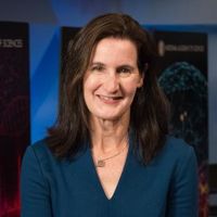
Dominique Bergmann

Joseph A Berry

Adrien Burlacot
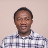
Barnabas Daru
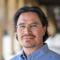
José R. Dinneny

Rodolfo Dirzo

David Ehrhardt
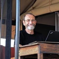
Chris Field
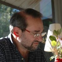
Arthur Grossman

Aidee Guzman

Andrew Leslie
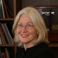
Sharon R. Long
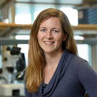
Erin Mordecai
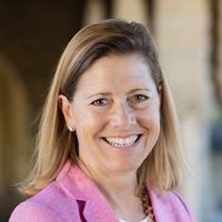
Mary Beth Mudgett
Virginia Walbot
| Content -->408 Panama Mall, Suite 217 Stanford, CA 94305-6032 United States . Stanford, California 94305. |
- Office of Graduate Education
Admissions Timeline
2025 -2026 admissions.
Mid-September 2024 Application Opens
December 1, 2024 Application Deadline (applying for KHS)
December 3, 2024 Application Deadline
Early-December, 2024 - Mid-January, 2025 Application Reviews
3rd week of January, 2025 Interview Invitations start to be sent
March 5 - March 8, 2025 Interview Session (in-person)
Mid-March, 2025 Admission offer letters sent
April 15, 2025 Deadline to accept admission offer
Matriculation

Welcome to the Hadly Lab
Our mission is to understand, describe, and predict how biodiversity responds to change in the Anthropocene.
We are a laboratory for life in the Anthropocene.
We envision a world that puts diversity first, in all living systems, wild and human. Just like biodiversity in the ecosystems we study, diversity in any system confers strength, resilience, and beauty. We actively bring together and integrate a diverse set of people, perspectives, scientific methods, tools, ecosystems and time-scales. Diversity statement and resources

Our research explores the ecology and evolution of animals. Using a variety of techniques, we sense patterns in nature to understand how the rapidly changing world of the Anthropocene is influencing the fate of biodiversity on Earth.

For us, teaching means learning together. Whether we are teaching, mentoring or communicating in a Stanford classroom or in Yellowstone, we seek to listen, understand, and create a shared vision of how healthy natural systems will support a thriving human society.

Data from genomes, the fossil record, camera traps, satellites, bat detectors and more allow us to sense the environment and the animals all around us - and how they respond to disturbance. We are developing novel field-ready technology and we are actively seeking collaborators.
Publications

What is the Anthropocene? Why are we concerned?
The world has reached a moment where human impact is as significant as the geologic forces of the past— this is called The Anthropocene. One million species are at risk of extinction, jeopardizing the natural world that makes our life possible and beautiful. We are studying these changes and communicating the findings so that we have the science and tools to better predict what's to come - and so we can all take action to create a future we can look forward to.


Elizabeth A Hadly
Principal investigator, dr. elizabeth hadly.
Elizabeth Hadly is a global change scientist who has studied the impacts of environmental change for the past four decades. Addressing the biology of species from both evolutionary and ecological perspectives, she studies primarily extant species to understand the past, present, and future of biodiversity. Uniquely, her research spans the decadal to millennial time scale, and integrates lab and field research, increasingly important in understanding the Anthropocene and tipping points for Planet Earth.
Read more about Liz
Meet the Team
Our current PhD students, postdocs and staff. Click on the link below to read about our undergraduates, community members and collaborators who are active and essential members of the Hadly Lab:
Hadly Lab Community and Alumni

Bryan Juarez

Jenni Rojas

Kirsten Verster

Tanvi Dutta Gupta
Jayela lopez.

Vrinda Suresh

M. Allison Stegner

Mary Ellen Hannibal
Research Teaching Tools Outreach
Welcoming Guolan Lu
We are excited to announce that Guolan Lu , PhD, has been appointed as an Assistant Professor at the Department of Urology.
Guolan is a biomedical engineer cross-trained in machine learning, biomedical imaging, and spatial omics in the field of cancer biology, immunology, and pharmacology. She completed her postdoctoral training at Stanford University in the laboratories of Dr. Eben Rosenthal, MD, and Dr. Garry Nolan, PhD, where she designed and led a first-in-human clinical study for fluorescence-guided surgery in pancreatic cancer, and pioneered a single-cell spatial pharmacology approach to decode drug-target-microenvironment interactions in clinical tumors. She received her Ph.D. from the joint Biomedical Engineering Department of Georgia Tech and Emory University. Her work has earned her multiple awards, including the K99/R00 Pathway to Independence Award from the NCI and recognition as a Rising Star in Biomedical Engineering (MIT). She was also a fellow in the Stanford Molecular Imaging Scholars (SMIS) Program and a Stanford Translational Medicine (TRAM) Scholar.

Guolan Lu Lab is dedicated to developing and integrating cutting-edge technologies in biomedical imaging, artificial intelligence, and spatial multi-omics to uncover the principles governing the spatiotemporal dynamics of tissue architecture and the mechanisms driving disease initiation, progression, and therapeutic response. Our ultimate goal is to translate this knowledge into next-generation theragnostics to cure both benign and malignant diseases.
In her free time, Guolan loves going on road trips, camping, and hiking with her family. She finds exploring nature together a great way to unwind and recharge.
Join us in welcoming Guolan!
Along with Stanford news and stories, show me:
- Student information
- Faculty/Staff information
We want to provide announcements, events, leadership messages and resources that are relevant to you. Your selection is stored in a browser cookie which you can remove at any time using “Clear all personalization” below.
Seeing what’s going on inside a body is never easy. While technologies like CT scans, X-rays, MRIs, and microscopy can provide insights, the images are rarely completely clear and can come with side effects like radiation exposure.
But what if you could apply a substance on the skin, much like a moisturizing cream, and make it transparent, without harming the tissue?
That’s what Stanford scientists have done using an FDA-approved dye that is commonly found in food, among several other light-absorbing molecules that exhibit similar effects. Published in Science on Sept. 5, the research details how rubbing a dye solution on the skin of a mouse in a lab allowed researchers to see, with the naked eye, through the skin to the internal organs, without making an incision. And, just as easily as the transparency happened, it could be reversed.
“As soon as we rinsed and massaged the skin with water, the effect was reversed within minutes,” said Guosong Hong , assistant professor of materials science and engineering and senior author on the paper. “It’s a stunning result.”
Absorption reduces scattering of light
When light waves strike the skin, the tissue scatters them, making it appear opaque and non-transparent to the eye. This scattering effect arises from the difference in the refractive indices of different tissue components, such as water and lipids. Water usually has a much lower refractive index than lipids in the visible spectrum, causing visible light to scatter as it goes through tissue containing both.
To match the refractive indices of different tissue components, the team massaged a solution of red tartrazine – also known as the food dye FD&C Yellow 5 – onto the abdomen, scalp, and hindlimb of a sedated mouse. The skin turned red in color, indicating that much of the blue light had been absorbed due to the presence of this light-absorbing molecule. This increase in absorption altered the refractive index of the water at a different wavelength – in this case, red. As a result of the absorption of the dye, the refractive index of water matches that of lipids in the red spectrum, leading to reduced scattering and making the skin appear more transparent at the red wavelength.
This research is a new application of decades-old equations that can describe the relationship between absorption and refractive index, called the Kramers-Kronig relations. In addition to this food dye, several other light-absorbing molecules have demonstrated similar effects, thereby confirming the generalizability of the underlying physics behind this phenomenon.
Researchers were able to see, without special equipment, the functioning internal organs, including the liver, small intestine, cecum, and bladder. They were also able to visualize blood flow in the brain and the fine structures of muscle fibers in the limb. The mouse’s beating heart and active respiratory system indicated that transparency was successfully achieved in live animals. Furthermore, the dye didn’t permanently alter the subject’s skin, and the transparency disappeared as soon as the dye was rinsed with water.
The researchers believe this is the first non-invasive approach to achieving visibility of a mouse’s living internal organs.
“Stanford is the perfect place for such a multifaceted project that brings together experts in materials science, neuroscience, biology, applied physics, and optics,” said Mark Brongersma , professor of materials science and engineering and co-author on the paper. “Each discipline comes with its own language. Guosong and I enjoyed taking each other’s courses on neuroscience and nanophotonics to better appreciate all the exciting opportunities.”
The potential future of ‘clear’ tissue
Right now, the study has only been conducted on an animal. If the same technique could be translated to humans, it could provide a range of biological, diagnostic, and even cosmetic benefits, Hong said.
For example, instead of through invasive biopsies, melanoma testing could be done by looking directly at a person’s tissue without removing it. This approach could potentially also replace some X-rays and CT scans, and make blood draws less painful by helping phlebotomists easily find veins. It could also improve services like laser tattoo removal by helping to focus laser beams precisely where the pigment is below the skin.
“This could have an impact on health care and prevent people from undergoing invasive kinds of testing,” said Hong. “If we could just look at what’s going on under the skin instead of cutting into it, or using radiation to get a less than clear look, we could change the way we see the human body.”
For more information
Other Stanford co-authors include Betty Cai, member of the Department of Materials Science Engineering ; Zihao Ou, Carl H. C. Keck, Shan Jiang, Kenneth Brinson Jr, Su Zhao, Elizabeth L. Schmidt, Xiang Wu, Fan Yang, Han Cui, and Shifu Wu, who are also with the Department of Materials Science Engineering and Wu Tsai Neurosciences Institute ; Yi-Shiou Duh of the Department of Physics and Geballe Laboratory for Advanced Materials ; Nicholas J. Rommelfanger of the Wu Tsai Neurosciences Institute and Department of Applied Physics ; Wei Qi and Xiaoke Chen of the Department of Biology ; Adarsh Tantry of the Wu Tsai Neurosciences Institute and Neurosciences IDP Graduate program ; Richard Roth of the Department of Neurosurgery ; Jun Ding of the Department of Neurosurgery and Department of Neurology and Neurological Sciences ; and Julia A. Kaltschmidt of the Wu Tsai Neurosciences Institute and Department of Neurosurgery .
This work was supported by the National Institutes of Health, National Science Foundation, Air Force, Beckman Technology, Rita Allen Foundation, Focused Ultrasound Foundation, Spinal Muscular Atrophy Foundation, Pinetops Foundation, Bio-X Initiative of Stanford University, Wu Tsai Neuroscience Institute, Knight-Hennessy, and U.S. Army Long Term Health Education and Training program.
Disclaimer: The technique described above has not been tested on humans. Dyes may be harmful. Always exercise caution when handling dyes – do not consume them, apply them to people or animals, or misuse them in any way.
Media contacts:
Jill Wu, School of Engineering: [email protected]

Guolan Lu, PhD
Principal Investigator
Guolan is an Assistant Professor in the Department of Urology at Stanford University School of Medicine. She is a biomedical engineer cross-trained in machine learning, biomedical imaging, and spatial omics in the context of cancer biology, immunology, and pharmacology. She completed her postdoctoral training in the laboratories of Dr. Garry Nolan, PhD and Dr. Eben Rosenthal, MD, at Stanford University, where she designed and led a first-in-human clinical study for fluorescence-guided surgery in pancreatic cancer, and pioneered a single-cell spatial pharmacology approach to decode drug-target-microenvironment interactions in situ in clinical tumors. She received her Ph.D. from the joint Biomedical Engineering Department of Georgia Tech and Emory University (advisor: Dr. Baowei Fei, PhD), where she received interdisciplinary training in medical imaging, machine learning, and digital pathology.
Guolan has received multiple awards for her work, including a K99/R00 Pathway to Independence Award from the National Cancer Institute, a Young Investigator Award from The Society for Immunotherapy of Cancer (SITC), and recognition as a Rising Star in Biomedical Engineering and Science (MIT), etc. She was a fellow of the Stanford Molecular Imaging Scholars (SMIS) Program and a Stanford Translational Medicine (TRAM) Scholar.

Eric Peterson, MBT, HTL
Lab Services Manager
Eric Peterson received his BS in biochemistry from UC Davis and a master's in biotechnology from San Jose State. He has worked in clinical laboratories for about 9 years, and in research labs for over 15. Those years were spent developing pharmaceuticals to treat coronary thrombosis, breast cancer, pain, and autoimmune disorders. Eric joined the School of Medicine in 2017 and moved to the Department of Urology in 2023. At SJSU, he teaches Biology 233T - Immunological Techniques in the Laboratory to the next generation of biotechnology grad students. In his spare time, he enjoys cooking and reading about history.

Ramon Borquez
Administrative Associate
Haleh Alimohamadi wins 2024 Spicer Young Investigator Award for breakthroughs in understanding cell membrane activity
Alimohamadi is being recognized for her novel integration of theoretical and experimental results to connect diverse health outcomes with cell membrane behavior.
By Kimberly Hickok
When Haleh Alimohamadi, a mechanical engineer by training, began talking to Gerard Wong about a postdoctoral position in his microbiology and immunology lab at University of California, Los Angeles, Wong was apprehensive. “I had never hired anyone who did not have previous experience doing experiments in my group,” he said.
Alimohamadi had a wealth of experience with computational modeling, but none when it came to experimentation. Despite his initial hesitation, Wong was impressed by Alimohamadi’s persistence and clear, creative ideas and took a chance on the rookie experimentalist, which he now believes “turned out to be one of the best things I did this decade.”
Now, as a bioengineering postdoctoral scholar in Wong’s group, Alimohamadi has leveraged small-angle X-ray scattering measurements at the Stanford Synchrotron Radiation Lightsource (SSRL) to describe in unprecedented scale and accuracy how the exterior membrane of the cells of living organisms responds to various proteins and peptides that come in contact with it. This information is crucial for understanding how pathogens, such as SARS-CoV-2, the virus responsible for COVID-19, infect and damage cells and cause widespread, lingering symptoms.
For her contributions, Alimohamadi has been awarded SSRL’s 2024 Spicer Young Investigator Award .
“I'm so thankful to the SSRL committee for choosing me,” Alimohamadi said. “It’s amazing to have the opportunity to do this work, especially for a scientist like me, whose background was in a completely different area.”
The shape of life
As a graduate student at the University of California, San Diego, Alimohamadi spent several years developing and analyzing computational models to understand biophysical phenomena at the nanoscale – specifically, how cell membranes can be deformed. But for her postdoc research, “I needed more,” she said. “I needed to manipulate something with my hands and see the events happen with my own eyes, in real life.”
Alimohamadi aimed to join a lab where she could experimentally validate her computational models. “I didn't have any background in doing experiments, neither in physics, nor in biology, but I joined Gerard Wong’s lab at UCLA, and he believed in me and gave me this opportunity,” she said. “That was a big turn in my life.”
Cell membrane-level experiments are at the nanoscale, meaning one-billionth of a meter – or smaller than one millionth of the thickness of a dime. Traditionally, experiments at this scale can be expensive, challenging, or destructive to the cell. For that reason, Alimohamadi aimed to combine biophysical models with SAXS measurements, which don’t destroy the cell.
The results allowed Alimohamadi and her colleagues to describe how the curvature of cell membranes changes in response to specific proteins. From there, the researchers identified the curvature-generating regions of proteins, such as those that are involved in cell death, division, or fusion processes. Understanding how these regions of proteins operate is important, Alimohamadi explained, because disruption in their function can cause several diseases, including cancer, neurodegenerative diseases, metabolic problems and more.
In under two years’ time working with Wong’s group, Alimohamadi has authored and contributed to more than 10 scientific publications in top-tier journals, including a first-authored study published in the Journal of the American Chemical Society in 2023, describing how proteins create pathways, or pores, through cell membranes. Earlier this year, she published another first-authored study in ACS Nano , describing how proteins induce membrane bending and separation processes.
“Learning to do the experiments has been challenging, but I love it,” Alimohamadi said. “This was a big turn in my life and I’m so happy that I took the risk.” As an immigrant, woman scientist, Alimohamadi said she is thankful to all of her mentors for their support.
She also hopes to encourage other scientists considering the shift from theoretical to experimental work to trust their instincts, and make the leap if they want to. “There are going to be several challenges, and you will fail several times, but it’s worth the risk,” she said. “Listen to yourself and do the work that makes you happy.”
The Spicer Award will be presented to Alimohamadi during the 2024 SSRL/LCLS Annual Users' Meeting at SLAC, which will be held September 22-27. The award is named in honor of the late William Spicer, who co-founded what would become SSRL, and his wife Diane.
SSRL is a DOE Office of Science user facility.
For questions or comments, contact Strategic Communications & External Affairs at [email protected] .
SLAC National Accelerator Laboratory explores how the universe works at the biggest, smallest and fastest scales and invents powerful tools used by researchers around the globe. As world leaders in ultrafast science and bold explorers of the physics of the universe, we forge new ground in understanding our origins and building a healthier and more sustainable future. Our discovery and innovation help develop new materials and chemical processes and open unprecedented views of the cosmos and life’s most delicate machinery. Building on more than 60 years of visionary research, we help shape the future by advancing areas such as quantum technology, scientific computing and the development of next-generation accelerators.
SLAC is operated by Stanford University for the U.S. Department of Energy’s Office of Science . The Office of Science is the single largest supporter of basic research in the physical sciences in the United States and is working to address some of the most pressing challenges of our time.
Related Topics
- Biological sciences
- X-ray scattering and diffraction
- X-ray science
- X-ray light sources and electron imaging
- Stanford Synchrotron Radiation Lightsource (SSRL)
Related stories
Scientists unlock the secrets of how a key protein converts dna into rna.
The results, which show how the protein adds nucleotides to the growing RNA chain, could lead to more effective medications.
SLAC and Stanford researchers advance understanding of a key celiac enzyme
Wheat and other sources of gluten can spell trouble for people with the disease, but new findings could aid the development of first-ever drugs...

A novel spray device helps researchers capture fast-moving cell processes
Researchers figured out how to spray and freeze a cell sample in its natural state in milliseconds, helping them capture basic biological processes in...
A newly published protein structure helps explain how some anti-cancer immunotherapy treatments work
Scientists at Stanford and NYU have published and investigated a new structure of the protein LAG-3 which could enable the development of new cancer...
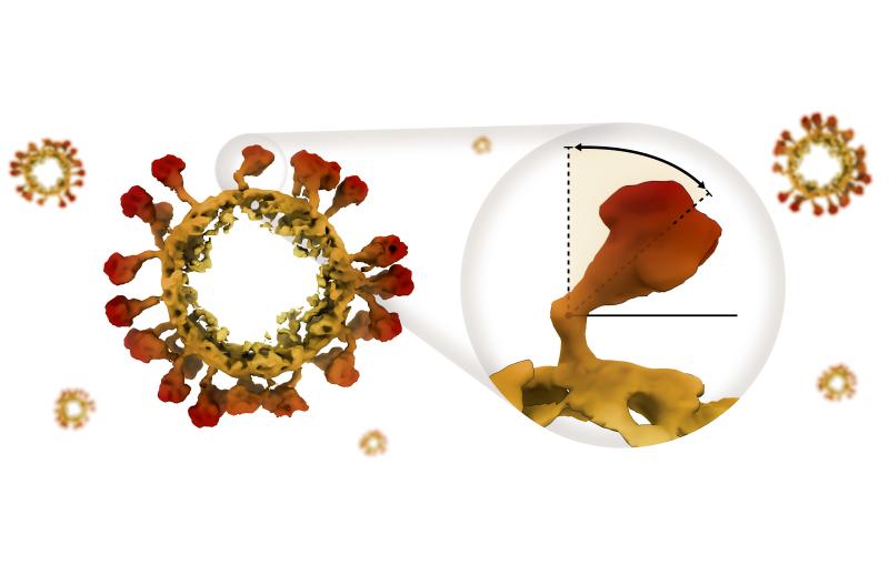
How tiny hinges bend the infection-spreading spikes of a coronavirus
Disabling those hinges could be a good strategy for designing vaccines and treatments against a broad range of coronavirus infections.
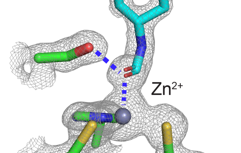
Stanford-SLAC study shows how modifying enzymes’ electric fields boosts their speed
A swap of metals and a mutation ramp up the electric field strength at the active site of an enzyme, making it ...

IMAGES
VIDEO
COMMENTS
The Biology Ph.D. program is part of the larger Biosciences community at Stanford, which includes doctorate programs in the basic science departments at Stanford Medical School. There are two tracks within the Biology Ph.D. program: Cell, Molecular and Organismal Biology. Ecology and Evolution. (Previously a part of the Department of Biology ...
Stanford Biology PhD Program applications are made through Graduate Admissions. The application deadline for Autumn Quarter 2024 matriculation is December 5, 2023 at 11:59pm pst. The application for the Autumn 2024 cohort will be available in September 2023. Please review the Graduate Admissions website prior to starting your application.
Welcome to the Biology Department! Our community is devoted to discovering fundamental knowledge of the living world: from the behavior of single molecules to dynamics of cells, organisms, populations, and interactions of biological systems with our planet. We are dedicated to developing innovative training experiences for the next generation ...
The Stanford Biology Preview Program (BPP) focuses on supporting scholars from diverse and non-traditional backgrounds in navigating the application process to PhD programs. The Stanford BPP offers virtual workshops to raise awareness about Stanford's Biology PhD Program application process, to build on the strengths of prospective PhD applicants, as well as provide individual mentorship and ...
(Previously a part of the Department of Biology Hopkins Marine Station is now a part of the Oceans Department within Stanford Doerr School of Sustainability) Some requirements are the same across concentrations, and other requirements are concentration-specific. Please review the Ph.D. Handbook for specific details.
How to Apply Thank you for your interest in the 14 Biosciences PhD Programs at Stanford University! The online application for Autumn 2024-25 will open in mid-September 2024.
Our 14 Biosciences PhD Home Programs empower students with the flexibility to tailor their education to their skills and interests as they evolve. Students work with global leaders in biomedical innovation, who provide the mentorship to answer the most difficult and important questions in biology and biomedicine. We encourage our students to flow freely between the 14 Biosciences PhD Home ...
Graduate Program in Stem Cell Biology & Regenerative Medicine Stanford is a world leader in stem cell research and regenerative medicine. Central discoveries in stem cell biology - tissue stem cells and their use for regenerative therapies, transdifferentiation into mature cell-types, isolation of cancerous stem cells, and stem cell signaling pathways - were made by Stanford faculty and ...
Chemical and systems biology of Ca2+ dependent phosphatase signaling networks Biochemistry Cell Biology Computational Biology Genetics
Program Overview. For graduate-level students, the department offers resources and experience learning from and working with world-renowned faculty involved in research on ecology, neurobiology, population biology, plant and animal physiology, biochemistry, immunology, cell and developmental biology, genetics, and molecular biology.
To be eligible for admission to graduate programs at Stanford, applicants must meet one of the following conditions: Applicants must hold, or expect to hold before enrollment at Stanford, a bachelor's degree from a U.S. college or university accredited by a regional accrediting association.
The Chemical and Systems Biology Ph.D. program also emphasizes collaborative learning, and our research community includes scientists trained in molecular biology, cell biology, chemistry, physics, and engineering. Our Ph.D. program consistently ranks among the top graduate training programs in the world. Most recently the National Research ...
The Stanford Structural Biology Graduate Home Program emphasizes research training, with coursework covering quantitative approaches to biology. Department research tackles biological problems at the atomic level, using structural and biophysical methodologies to explain both function and disease.
Cancer Biology PhD Program. Established in 1978, the interdisciplinary Cancer Biology PhD Program is designed to provide graduate and medical students with the education and training they need to make significant contributions to the field of cancer biology. The program is led by Laura Attardi, PhD, and Julien Sage, PhD, and currently has over ...
Biosciences PhD students began their careers at Stanford School of Medicine with crisp new lab coats, advice on graduate school success and warm words about the value of discovery.
Prospective PhD Students Coterm Students Postdoctoral Scholars Administrative Staff Faculty Affiliated Programs Bio-X CEHG ChEM-H Hopkins Marine Station Woods Institute Wu Tsai Neuro Contact Us Gilbert Building 371 Jane Stanford Way Stanford, CA 94305 Phone: 650-723-2413 [email protected] Campus Map SUNet Login Stanford University ...
PhD Program Understanding Cell Signalling is a unifying theme in The Department of Molecular and Cellular Physiology (MCP). Our PhD program engages graduate students in research across multiple disciplines - including structural biology, biophysics, cell biology, and neuroscience in a highly collaborative environment. Our faculty, who teach courses in physiology, cell biology, neuroscience ...
The Cancer Biology Program welcomes graduate applications from individuals with a broad range of life experiences, perspectives, and backgrounds who would contribute to our community of scholars. The review process is holistic and individualized, considering each applicant's academic record and accomplishments, letters of recommendation, prior research experience, and admissions essays to ...
This new thrust in developmental biology derives from the extraordinary methodological advances of the past decade in molecular genetics, immunology, and biochemistry. However, it also derives from groundwork from classical developmental studies, the rapid advances in cell biology and animal virology, and models borrowed from prokaryotic systems.
The Stanford Biosciences is an umbrella program that provides a single application and unified graduate training across a range of disciplines in biology and biomedical research. The Biosciences program encourages students to explore research opportunities, carry out rotations, and choose dissertation research in any of the 14 Home Programs.
2025 -2026 Admissions. Mid-September 2024 Application Opens. December 1, 2024 Application Deadline (applying for KHS) December 3, 2024 Application Deadline. Early-December, 2024 - Mid-January, 2025 Application Reviews. 3rd week of January, 2025 Interview Invitations start to be sent. March 5 - March 8, 2025 Interview Session (in-person)
The mission of the Cancer Biology PhD program is to train graduate students so that they may ultimately launch careers related to the study and treatment of cancer. A major goal of program is to assist students in their growth and development by constructing meaningful educational plans. We believe that students will become outstanding cancer ...
Elizabeth Hadly is a global change scientist who has studied the impacts of environmental change for the past four decades. Addressing the biology of species from both evolutionary and ecological perspectives, she studies primarily extant species to understand the past, present, and future of biodiversity.
She completed her postdoctoral training at Stanford University in the laboratories of Dr. Eben Rosenthal, MD, and Dr. Garry Nolan, PhD, where she designed and led a first-in-human clinical study for fluorescence-guided surgery in pancreatic cancer, and pioneered a single-cell spatial pharmacology approach to decode drug-target-microenvironment ...
It's why he applied to the BioRETs INSPIRE program at Stanford, a collaboration between the GSE's Center to Support Excellence in Teaching (CSET) and the Department of Biology at the Stanford School of Humanities and Sciences. The program, supported by the U.S. National Science Foundation (NSF), connects Bay Area middle and high school ...
"Stanford is the perfect place for such a multifaceted project that brings together experts in materials science, neuroscience, biology, applied physics, and optics," said Mark Brongersma ...
Guolan Lu, PhD. Principal Investigator. [email protected]. Guolan is an Assistant Professor in the Department of Urology at Stanford University School of Medicine. She is a biomedical engineer cross-trained in machine learning, biomedical imaging, and spatial omics in the context of cancer biology, immunology, and pharmacology.
As a graduate student at the University of California, San Diego, Alimohamadi spent several years developing and analyzing computational models to understand biophysical phenomena at the nanoscale - specifically, how cell membranes can be deformed. But for her postdoc research, "I needed more," she said.